44 onion cells under microscope with labels
› 44090147 › CambridgeCambridge International AS and A Level Biology Coursebook ... Enter the email address you signed up with and we'll email you a reset link. Animal Cell Under Light Microscope Labelled : Draw and label the ... Onion cell diagram labeled structure of animal cell and plant cell under microscope. An organelle found in large numbers in most cells, in which the biochemical processes of respiration and energy production occur. Under a light microscope, the cell membrane, nucleus and cytoplasm of a cheek cell (animal cell) can be observed.
Onion Cell Under Microscope 400x Labeled - Micropedia Onion Epidermis. Px12 016a Plant Stomata Guard Cells Opened Zebrina Spp 400x. Onion Cells Under The Microscope Requirements Preparation And. Microscopy Biology Libretexts. Cells Under A Microscope By Jaimarie Nelson. Cell Lab. Microscope Lc Lessons Tes Teach. Human Cheek Cell Hamle Rsd7 Org.

Onion cells under microscope with labels
Onion Cell Microscope Slide Experiment - YouTube -- Materials: A Microscope, A Microscope Camera*, Microscope Slides, Coverslips, A Kitchen Knife, Pipettes*, Iodine Tincture, An Onion, Cutting Board -- I wrote a book about STEM! STEM Files is a... PDF Onion Cells - Investigation - Exploring Nature 5. Observe the onion tissue under the microscope at 4x, 10x and 40x with lots of light (open diaphragm). Then slowly close the diaphragm while observing the image to find the best light for seeing cellular details. 6. Draw a section of onion skin cells at 10x magnification. Then switch to 40x and draw one cell and label it. Questions: 1. › natural-sciences › gr9Natural Sciences Grade 9 - Grade 7-9 Workbooks The onion cells have a thick cell wall and a cell membrane. The animal cells only have a cell membrane. The onion cells have a regular shape whereas the cheek cells have a irregular shape and seem more flimsy. In the onion cells they might notice a large vacuole which might not be as visible in the cheek cells. Cheek cells do not have vacuoles ...
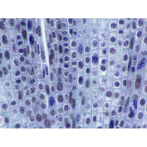
Onion cells under microscope with labels. sciencequiz.net › newjcscience › jcbiologyThe Cell - ScienceQuiz.net The diagram shows a group of onion cells. The parts labelled A, B and C respectively are ... The diagram shows a plant cell as seen under a microscope. Two of the ... VIEWING PLANT CELLS UNDER THE MICROSCOPE: onion ... This method allows students to view plant cells under the microscope. A single layer of onion cells (membrane) can be easily obtained from the bulb. These can ...2 pages Onion Cells Under a Microscope - Requirements/Preparation/Observation Add a drop of iodine solution on the onion membrane (or methylene blue) Gently lay a microscopic cover slip on the membrane and press it down gently using a needle to remove air bubbles. Touch a blotting paper on one side of the slide to drain excess iodine/water solution, Place the slide on the microscope stage under low power to observe. Onion Root Cells Dividing By Mitosis Under A Light Microscope ... - iStock Download this Onion Root Cells Dividing By Mitosis Under A Light Microscope At 100x Magnification Cells Visible In Prophase Metaphase Anaphase And Telophase photo now. And search more of iStock's library of royalty-free stock images that features Anaphase photos available for quick and easy download.
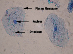
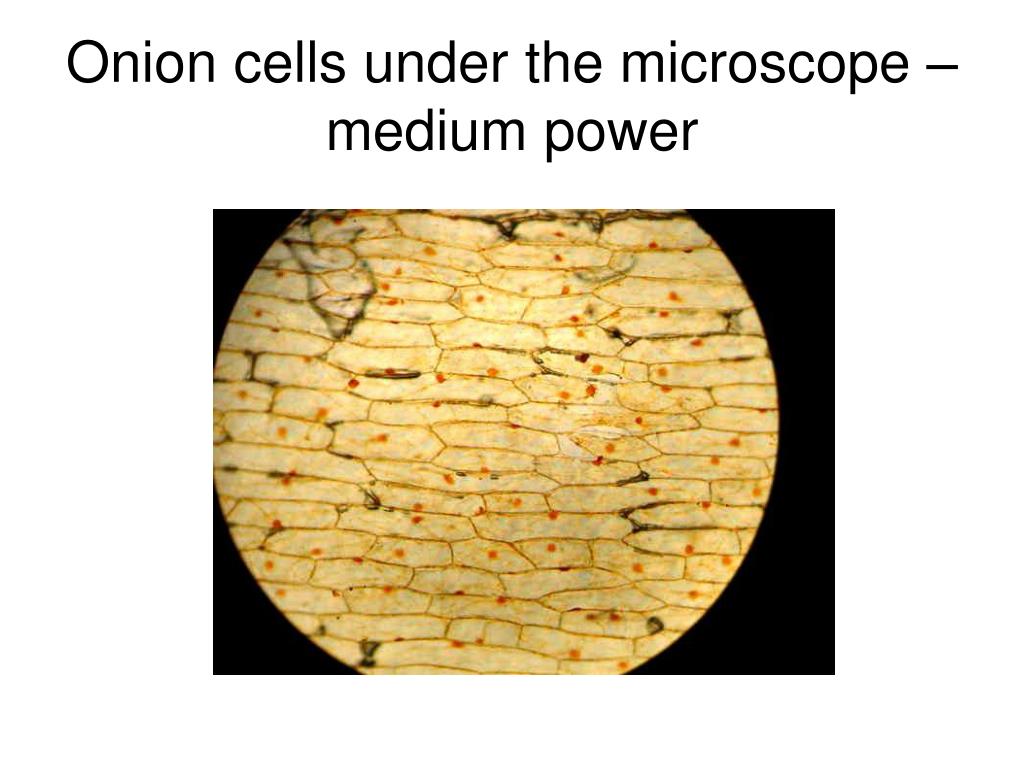
Cheek Cells Under a Microscope - Requirements/Preparation/Staining The nucleus at the central part of the cheek cell contains DNA. When a drop of methylene blue is introduced, the nucleus is stained, which makes it stand out and be clearly seen under the microscope. Although the entire cell appears light blue in color, the nucleus at the central part of the cell is much darker, which allows it to be identified. Onion cells under the microscope: 40X - 100X - 400X - YouTube under the #microscope: 40X - 100X - 400X Onion Cells Microscope Stock Photos and Images - Alamy Onion Cells under the Microscope ID: FC0CWP (RF) Onion skin cells under the microscope, horizontal field of view is about 0.61 mm ID: 2AM97C0 (RM) Detailed view of the cells of a red onion as seen through a microscope. Biology experiment. ID: 2H3C0XC (RF) Onion cells under a light microscope at 10 times magnification ID: 2EW4298 (RF) › bitesize › articlesCells and Reproduction - BBC Bitesize Onion cells are easy to see using a light microscope. ... A small tube placed under the skin of the upper arm. ... Five small tubes with labels and stoppers or lids Cress seeds Labels Cotton wool ...
Observing Onion Cells Under The Microscope Afterwards, carefully mount the prepared and stained onion cell slide onto the microscope stage. Make sure that the cover slip is perfectly aligned with the microscope slide, and that any excess stain has been wiped off. Secure the slide on the stage using the stage clips. Using a light microscope - Required Practical Review Biology Practical - Using a light microscope to observe, draw and label cells in an onion skin. Video link: ...8 pages Animal Cell Diagram Under Microscope Labeled Animal Cell Diagram Under Microscope. Function cell does in the body dictate the change and adaptation done by cell. When observing onion cells, there is the Cell Surface Membrane which is present in all living cells. We all keep in mind that the human body is quite intricate and a method I discovered to are aware of it is via the manner of ... Microscopy, size and magnification - Microscopy, size and ... - BBC Place cells on a microscope slide. Add a drop of water or iodine (a chemical stain). Lower a coverslip onto the onion cells using forceps or a mounted needle. This needs to be done gently to...
Microscope Cell Lab: Cheek, Onion, Zebrina - SchoolWorkHelper The first lab exercise was observing animal cells, in this case, my cheek cells. The second lab exercise was observing plant cells, in this case, onion epidermis. The third lab exercise was observing chloroplasts and biological crystals, in this case, a thin section from the Zebrina plant. The first thing that was done in this lab exercise was ...
Blog, She Wrote - Embracing the Independent & Authentic Nature of ... Blog, She Wrote - Embracing the Independent & Authentic Nature of ...
Onion Epidermal Cell Labeled Diagram - schematron.org Draw a labelled diagram of an onion epidermal cell seen under the microscope. ( 4 marks) e The onion epidermal cells are not green in colour because they lack. The epidermal cells of onions provide a protective layer against viruses and fungi that may harm the sensitive tissues.
Labeled Onion Cell Under Microscope 40x - Micropedia For the experiment you will only need onion dropper and the microscope container and tools are optional. Labeled onion cell under microscope 40x. While photosynthesis takes place in the leaves of an onion containing chloroplast the little glucose that is produced from this process is converted in to starch starch granules and stored in the bulb.
Onion Skin Cells - Investigation - Exploring Nature 5. Observe the onion tissue under the microscope at 4x, 10x and 40x with lots of light (open diaphragm). Then slowly close the diaphragm while observing the image to find the best light for seeing cellular details. 6. Draw a section of onion skin cells at 10x magnification. Then switch to 40x and draw one cell and label it.
Looking at the Structure of Cells in the Microscope Both types of light microscopy are widely used to visualize living cells. Figure 9-7 Two ways to obtain contrast in light microscopy. (A) The stained portions of the cell reduce the amplitude of light waves of particular wavelengths passing through them. A colored image of the cell is thereby obtained that is visible in the ordinary way. (more...)
pradeepresearch.org › publicationsPublications – Pradeep Research Group Facile crystallization of ice I h via formaldehyde hydrate in ultrahigh vacuum under cryogenic conditions, Jyotirmoy Ghosh, Gaurav Vishwakarma, Subhadip Das, and Thalappil Pradeep, J. Phys. Chem. C, 125 (2021) 4532–4539 (DOI: 10.1021/acs.jpcc.0c10367). PDF File Supporting Information
ocr.org.uk › Images › 643844-question-paper-depth-inOxford Cambridge and RSA Friday 16 October 2020 – Morning 1 (a) A student was observing onion epithelial cells using a light microscope. They photographed these cells and the image obtained is shown in Fig. 1.1. The student then made a drawing of a few cells from this image. The drawing is shown in Fig. 1.2. Fig. 1.1 cytoplasm cell wall large permanent vacuole ribosome Fig. 1.2
Biology Project The Biology Project, an interactive online resource for learning biology developed at The University of Arizona. The Biology Project is fun, richly illustrated, and tested on 1000s of students.
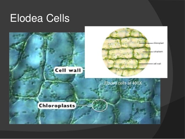
Plant Cell Under Microscope 40X Labeled : 1 - Chloroplast and cell wall ... The different images below were taken with two different types of microscopes. 1.can only turn fine adjustment 2.draw one row of cells across the middle 3.label the chloroplasts and cell wall. When using the microscope always start by focusing under low power and working your way up to high power.
Observing Cork Cells Under The Microscope Method 1. To start, prepare a wet mount by placing a tiny water droplet on the center of a clean microscope slide. Using a wet mount will keep the cork sample in place instead of sliding or flying off of the slide. Then, dip your finger inside the cork container to pick up some dust or shavings.
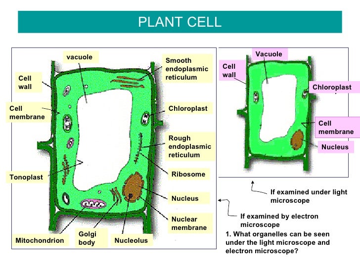
Plant Cell Under Microscope 40X Labeled - Powerpoint Lab Comparing ... The bulb of an onion is formed from modified leaves. Put a drop of water on the microscope slides. Unlike most plant cells, this species do not have a cell wall. See how a generalized structure of an animal cell and plant cell look with labeled diagrams. Under the microscope, plant cells are seen as large rectangular interlocking blocks.
› natural-sciences › gr9Natural Sciences Grade 9 - Grade 7-9 Workbooks The onion cells have a thick cell wall and a cell membrane. The animal cells only have a cell membrane. The onion cells have a regular shape whereas the cheek cells have a irregular shape and seem more flimsy. In the onion cells they might notice a large vacuole which might not be as visible in the cheek cells. Cheek cells do not have vacuoles ...
PDF Onion Cells - Investigation - Exploring Nature 5. Observe the onion tissue under the microscope at 4x, 10x and 40x with lots of light (open diaphragm). Then slowly close the diaphragm while observing the image to find the best light for seeing cellular details. 6. Draw a section of onion skin cells at 10x magnification. Then switch to 40x and draw one cell and label it. Questions: 1.
Onion Cell Microscope Slide Experiment - YouTube -- Materials: A Microscope, A Microscope Camera*, Microscope Slides, Coverslips, A Kitchen Knife, Pipettes*, Iodine Tincture, An Onion, Cutting Board -- I wrote a book about STEM! STEM Files is a...







)

Post a Comment for "44 onion cells under microscope with labels"